|
|
MAIN
KEYNOTE SPEAKER:
|
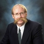
John C. Gore, Ph.D.
Chancellor University Professor of Radiology
and Radiological Sciences, Biomedical Engineering, and
Physics
Director, Vanderbilt University Institute of
Imaging Science
Department of Radiology and Radiological
Sciences
Profile
Dr. John Gore is Professor of Radiology and
Radiological Sciences, Biomedical Engineering, Molecular
Physiology and Biophysics, and Physics, and Director of the
Center for Imaging Sciences at Vanderbilt University Medical
Center. He is an international expert in the field of MRI
research.
Research Specialty:
Imaging Science, Magnetic Resonance Imaging (MRI)
|
 |
|
Research Description |
Dr. Gore's research program is focused on the
development and application of imaging, especially magnetic
resonance imaging and spectroscopy techniques, in clinical and basic
science. Imaging of human subjects and small animals provides unique
information on tissue structure and function, and is being applied
in a variety of different applications in neuroscience, cancer
research and studies of metabolism. A general theme of interest is
to understand the physical and physiological factors that affect MRI
signals and to use this knowledge to devise non-invasive imaging
methods that provide new types of information as well as for
developing new applications of imaging. A second major theme is the
development of methods for studying human brain structure and
function using MRI and for integrating fMRI data with other imaging
methods such as NIR and EEG. Applications of structural and
functional MRI to the brain are performed in collaboration with
investigators from psychology, psychiatry and other departments. A
third major theme is the use of multi-modality imaging (MRI, PET,
CT, optical and ultrasound) to study small animals, including mouse
models of human cancer and other genetically modified mice. Many
projects also involve the development and application of advanced
image analysis methods and computer algorithms. |
|
Keynote title:
Challenges and Opportunities
for High Field MRI
and MRS |
|
Abstract |
Magnetic resonance
imaging (MRI) and spectroscopy (MRS) have contributed considerably
to our understanding of the structure and function of the human
brain. For example, they have been used to evaluate the anatomic and
functional changes that occur with development or as a result of
aging, genetic or pathological influences. In recent years several
technical and conceptual advances have been made which promise to
increase the role of neuroimaging methods in both research and
clinical applications. In particular, very high field structural MRI,
functional MRI (fMRI) and MRS, as well as advanced methods of image
analysis, have considerable potential for increasing our
understanding of brain architecture, activity and metabolism. The
development and applications to animal brains and human subjects of
MRI systems at fields of 7 Tesla and above provide many new
opportunities and technical challenges. The increased signal
strength at higher fields enables higher resolution images to be
acquired, which provides new insights into the structural anatomy of
the brain, including higher resolution tractography of white matter.
These benefits have already been realized in several studies of
brain structure and function including functional studies of neural
connectivity and the effects of pharmacological agents. However,
high field MR imaging is also affected by macroscopic field
variations caused by inhomogeneities of magnetic susceptibility
within the body, and these can degrade spectra and introduce image
distortions. Moreover, the performance of radiofrequency (RF) coils
is also affected at higher fields, and it is more difficult to
create uniform RF fields within large objects. These challenges are
being met using various technical innovations such as dynamic
shimming, the use of parallel arrays of coils, novel
spectral-spatial excitation methods, novel pulse sequences and
post-acquisition digital processing. In combination these efforts
promise to allow ultra-high field imaging and spectroscopy to
contribute significantly to our understanding of brain structure and
function in various conditions.
|
SPECIAL SUBJECT
KEYNOTE SPEAKER 1:
|
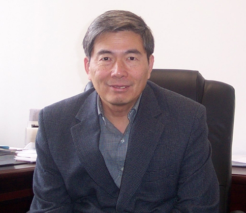
Dr. Robert E. Burrell
Professor and Chair
Department of Biomedical Engineering
Faculties of Engineering and of Medicine &
Dentistry
Professor and Canada Research Chair in Nanostructured
Biomaterials
Department of Chemical and Materials Engineering
Faculty of Engineering, University of Alberta |
 |
Profile
Dr. Robert
E. Burrell is currently a Canada Research Chair in Nanostructured
Biomaterials and a Professor of Chemical and Materials Engineering
in the Faculty of Engineering as well as Professor and Chair of
Biomedical Engineering in the Faculty of Medicine and Dentistry at
the University of Alberta. He is the past Vice President, Science
and Technology and Chief Scientific Officer, for Nucryst
Pharmaceutical Corp. He has a B.Sc. in Zoology and a M.Sc. in
Microbiology, University of Guelph and a Ph.D. in Microbiology,
University of Waterloo. He also held a Post Doctoral Fellowship in
Chemical Engineering at the University of Waterloo. He is an
Adjunct Professor at the University of Calgary. Prior to joining
Westaim, Dr. Burrell established a program for the development of
advanced industrial materials for biomedical and biotechnical
applications at Alcan International.
Research Specialty
Dr. Burrell is one of the world’s leading experts on
the use of advanced metallic films for therapeutic applications
including; 1) the control of microbial growth on a wide range of
devices and 2) control of the inflammatory response after injury.
He is the inventor of various devices and processes, including
antimicrobial films (Acticoat dressings, the worlds first commercial
medical application of nanotechnology to therapeutic treatments),
visual immunoassays (the application of nanotechnology to
diagnostics), submicron superparamagnetic particles (the application
of nanotechnology to cell sorting and separation), and single cell
protein production, for which 280 patents and pending patents exist
worldwide.
|
Research Description |
Dr. Robert
Burrell developed what is believed to be the world’s first
commercial medical application of nanotechnology – a bandage that
has revolutionized wound care and saved the lives and limbs of
patients around the world, including victims of the terrorist
attacks in both New York and Bali.
Wound dressings using Dr. Burrell’s patented
silver-based nanostructured biomaterials not only have
anti-microbial and anti-inflammatory properties but also speed
healing. The products are manufactured in Fort Saskatchewan under
the Acticoat brand by Nucryst Pharmaceuticals (formerly Westaim
Biomedical Inc.) and marketed by Smith & Nephew PLC, the global
leader in wound care.
Dr. Burrell began developing nanotechnology-based
processes and products to solve specific problems in medicine and
biotechnology while working in industry in the mid-1980z, before
nanotechnology became a common word among scientists. He is named
as the inventor on more than 30 U.S. patents and patent applications
and is considered to be the world authority on using silver to
improve wound healing.
While silver has been used for medicinal purposes for
thousands of years, development of an effective, stable and
convenient form of silver for wound treatment had eluded
scientists. Dr. Burrell’s invention of the nanocrystalline silver
technology on which Acticoat is based was a significant scientific,
clinical and commercial breakthrough.
Dr. Burrell developed the first strategic business
plan for Westaim Biomedical Inc. and as Chief Scientific Officer
developed an interdisciplinary team that brought eight new wound
care products, including Acticoat, to market in the first six
years. Before accepting a professorship at the University of
Alberta, Dr. Burrell initiated the connection that led Smith &
Nephew to acquire worldwide commercial rights to the Acticoat
product and technology in 2001. The business in worth $33 million
worldwide. Production at Fort Saskatchewan is expected to double
over the next two to three years.
Acticoat has been used by the University of Alberta
Hospital Burn Unit and to treat home care patients in Calgary. The
technology has the potential for application in other conditions
where infection or inflammation is a contributing factor, including
diabetic foot ulcers, psoriasis, rheumatoid arthritis and severe
chronic wounds. The combined would care and pharmaceutical value of
nanocrystalline technology is anticipated to be more than $500
million.
Dr. Burrell has been honored with a Canada Research
Chair and is in demand worldwide as a speaker and consultant. His
efforts clearly demonstrate that world-leading technology can be
successfully developed and commercialized in Alberta.
|
Keynote title: New
Frontiers in Biomedical Engineering: Nano and Regenerative Medicine
|
Abstract |
There is a considerable need for appropriate
infrastructure and expertise to successfully undertake
research in nano and regenerative medicine. The Faculty of
Engineering at the University of Alberta is one of the
highest rated research schools of engineering in North
America. It recently joined with the Faculty of Medicine
and Dentistry to create a dual faculty Department of
Biomedical Engineering. Further, the University of Alberta
is also the host site for the National Research Councils,
National Institute for Nanotechnology. Together this has
created the critical mass for research in nano and
regenerative medicine. Research is being conducted on drug
delivery using nanopolymers and aerosols, bone and tissue
regeneration and diagnostics including MEMS and NEMS
devices. My own research is on the use of nanostructured
noble metals (e.g. silver) for dermal tissue regeneration.
Historically, the use of silver as a medical
treatment dates to the 1880s when Crede used a 1% silver
nitrate solution to treat and prevent ophthalmia neonatorum.
Von Behring soon demonstrated that dilute forms of silver
nitrate were effective against typhoid and anthrax bacilli.
It was the activity of the dilute forms of silver nitrate
that led von Nägeli to coin the term oligodynamic activity.
Through the middle 20th century, silver fell into
disuse as the medical world concentrated on antibiotics for
the control of infection. The sixties saw a resurgence in
silver usage as burn physicians searched for more effective
broad spectrum antimicrobial agents. Silver nitrate (0.5%
solution) and silver sulfadiazine cream (1%) became the
standard forms of silver used in burn treatment. These
forms of silver all released only one form of silver, the
single valent silver ion (Ag+). It was in the
middle 1990s that nanotechnology created the opportunity to
develop a new delivery system that released biologically
active species other than Ag+. This technical
innovation has changed the spectrum of biological activity
found in silver releasing products. The single valent
silver ion was noted only as an antimicrobial agent and when
it was delivered from a cream or nitrate it was thought to
be pro-inflammatory and potentially detrimental to wound
healing. Nanocrystalline silver releases multiple species
of silver into solution and these new species have very
different biological properties than Ag+. It has
clearly been demonstrated in vitro, in vivo
and clinically that this new form of silver has unique
anti-inflammatory and antimicrobial properties.
In this presentation I will discuss: 1) The
infrastructure necessary for a successful program in nano
and regenerative medicine; and 2) The development of
nanocrystalline silver as a wound care product and its
unique physical, chemical and biological properties. This
will include its effects on the inflammatory and immune
systems using histological and immunohistochemical data
which result in regeneration of dermal tissue. |
SPECIAL SUBJECT
KEYNOTE SPEAKER 2:
|
K. Kirk Shung
Director and
Principal Investigator
Resource Center for Medical Ultrasonic Transducer Technology
Professor, Department of Biomedical Engineering
University of Southern California
http://bme.usc.edu/UTRC/ |
 |
Research Specialty:
Professor Shung currently conducts research primarily
in the area of high frequency ultrasonic imaging and transducer
development supported by an NIH National Resource grant on
transducer technology. Its thrust is on developing high frequency
ultrasonic transducers and arrays which with improved spatial
resolution are and can be used for dermatological, ophthalmological
and intravascular imaging and small animal imaging.
Keynote title: Medical
Ultrasound: Past, Present,
and
Future
|
Abstract |
Ultrasound
has been used as a diagnostic tool for more than 40 years.
Many medical applications have been found, mostly notably in
obstetrics and cardiology. It had a humble beginning,
started by a few curious scientists and clinicians in
different parts of the world in early 1950s and did not
become an established diagnostic tool until early 1970s when
grey scale ultrasonography was introduced. Modern ultrasound
scanners are capable of producing images of anatomical
structures in great detail in grey scale and of blood flow
in a color scale in real-time. State of the art 4D scanners
yielding 3D volumetric images in real-time are pushing the
technical envelope further. Today ultrasound is the
second-most used clinical imaging modality next only to
conventional x-ray radiography.
Although
ultrasound is considered to be a mature technology,
technical advances are still constantly being made. The most
significant achievements in ultrasound recently have been in
the developments of approaches capable of quantitative
measurement of tissue elastic properties, namely ultrasound
elastography and radiation force imaging, high frequency
imaging yielding improved spatial resolution and therapeutic
applications in drug delivery and high intensity focused
ultrasound surgery.
In this
talk, the history and current state of medical ultrasound
will be reviewed and future developments discussed. |
SPECIAL SUBJECT KEYNOTE
SPEAKER 3:
|
Regis B. Kelly, Ph.D.
Director, the
California Institute for Quantitative Biosciences (QB3)
Regis B. Kelly, Ph.D. is a distinguished
neuroscientist and former executive vice chancellor of UCSF
(from October, 2001 until January 31, 2004). As executive
vice chancellor, Kelly oversaw the UCSF research enterprise,
which now totals about $465 million annually. He also forged
new research ties between the university and private
industry. Kelly directs the institute at its headquarters on
UCSF's Mission Bay campus and also maintains a Berkeley
office. |
 |
Keynote title:
Synthetic Biology: Solving Biological Problems using
Engineering Principles
|
Abstract |
Biology, like astronomy and geology, has long
been a descriptive science. The challenge is to turn biology
into an engineering science where we design, build and test
solutions to important problems but using biological
materials. An early step has been to create a repository of
standardized parts that can be simply connected to perform
simple tasks, such as act as a photographic film, or an edge
detector. The next step is to use these skills to solve
problems, such as generating high octane fuel from waste
biological material. Applying engineering principles to
biology has opened up new possibilities, using
nanotechnology, microfluidics and advanced optics to monitor
the environment and to detect disease before symptom appear. |
|


